tree in bud opacities treatment
Radiation pneumonitis is the acute manifestation of radiation-induced lung disease and is relatively common following radiotherapy for chest wall or intrathoracic malignancies. A few COVID-19 cases and findings by dataset.

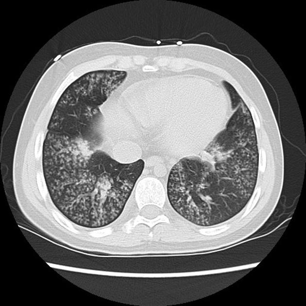
Tree In Bud Sign Lung Radiology Reference Article Radiopaedia Org
Pleural effusions are also more frequently encountered in this group of patients.
. A cystic form of Pneumocystis pneumonia is also. When compared to pulmonary tuberculosis upper lobe cavitation is less common and middle lobe bronchiectasis more frequent in Mycobacterium avium complex pulmonary infections. His chest CT images showed multi-focal patchy ground-glass opacities and parenchymal consolidation with both ill- and well-defined opacities predominantly involving the peripheral and posterior regions of both lungs.
A high-resolution CT HRCT is more sensitive to changes such as bronchiectasis small nodules tree-in-bud appearance ground glass opacities and pleural thickening. Coronal reconstructed computed tomography image shows the lingular cavity with irregular nodules and right mid-lung nodular opacities in a 43-year-old man who presented with cough and hemoptysis same patient as above. A aachen aardvark aardvarks aaron aba ababa abaci aback abactor abactors abacus abacuses abaft abalone abandon abandoned abandonee abandonees abandoning abandonment.
Please refer to the article on radiation-induced lung disease for a general discussion and radiation-induced pulmonary. Small patchy peripheral opacities are also present in the left lower lobe. Appearances typical of COVID-19 Figure Figure7.
This article does not deal with the changes seen in the late phase. Small nodules and tree-in-bud opacities are uncommon in patients with AIDS and pneumocystis pneumonia and usually indicate the presence of intercurrent infectious bronchiolitis from other organisms. In the right mid-lung nodular opacities are in a tree-in-bud distribution suggestive of endobronchial spread.
Coronal reconstructed computed tomography image shows the lingular. A Cardio-vasal shadow within the limits b Increasing left basilar opacity is visible arousing concern about pneumonia c Progressive infiltrate and consolidation d Small consolidation in right upper lobe and ground-glass opacities in both lower lobes e Infection demonstrates right infrahilar airspace. Treatment with tumor necrosis factoralpha TNF-α antagonists which is used for rheumatoid arthritis psoriasis.
He unusually also had a focal area of tree-in-bud opacification in the right lower lobe. In the right mid-lung nodular opacities are in a tree-in-bud distribution suggestive of endobronchial spread.
2

Chest Ct With Multifocal Tree In Bud Opacities Diffuse Bronchiectasis Download Scientific Diagram
View Of Tree In Bud The Southwest Respiratory And Critical Care Chronicles
2

Tree In Bud Pattern Pulmonary Tb Eurorad
2
View Of Tree In Bud The Southwest Respiratory And Critical Care Chronicles

Tree In Bud Sign Lung Radiology Reference Article Radiopaedia Org
2
Infectious Bronchiolitis With Extensive Tree In Bud Pattern Radiology Case Radiopaedia Org

Pdf Tree In Bud Semantic Scholar

References In Causes And Imaging Patterns Of Tree In Bud Opacities Chest

Pdf Tree In Bud
2

Hrct Scan Of The Chest Showing Diffuse Micronodules And Tree In Bud Download Scientific Diagram

References In Causes And Imaging Patterns Of Tree In Bud Opacities Chest

D E Lung Ct Shows Decrease In Bronchiectasis Ground Glass And Tree In Download Scientific Diagram
2

Computed Tomography Of The Chest Showed Nodular Opacities With Tree In Download Scientific Diagram